Fluorescence Molecular Imaging Systems were firstly introduced by Garofalakis and his colleagues. The first generation of this imaging system was designed for Non-contact Fluorescence Molecular Tomography.
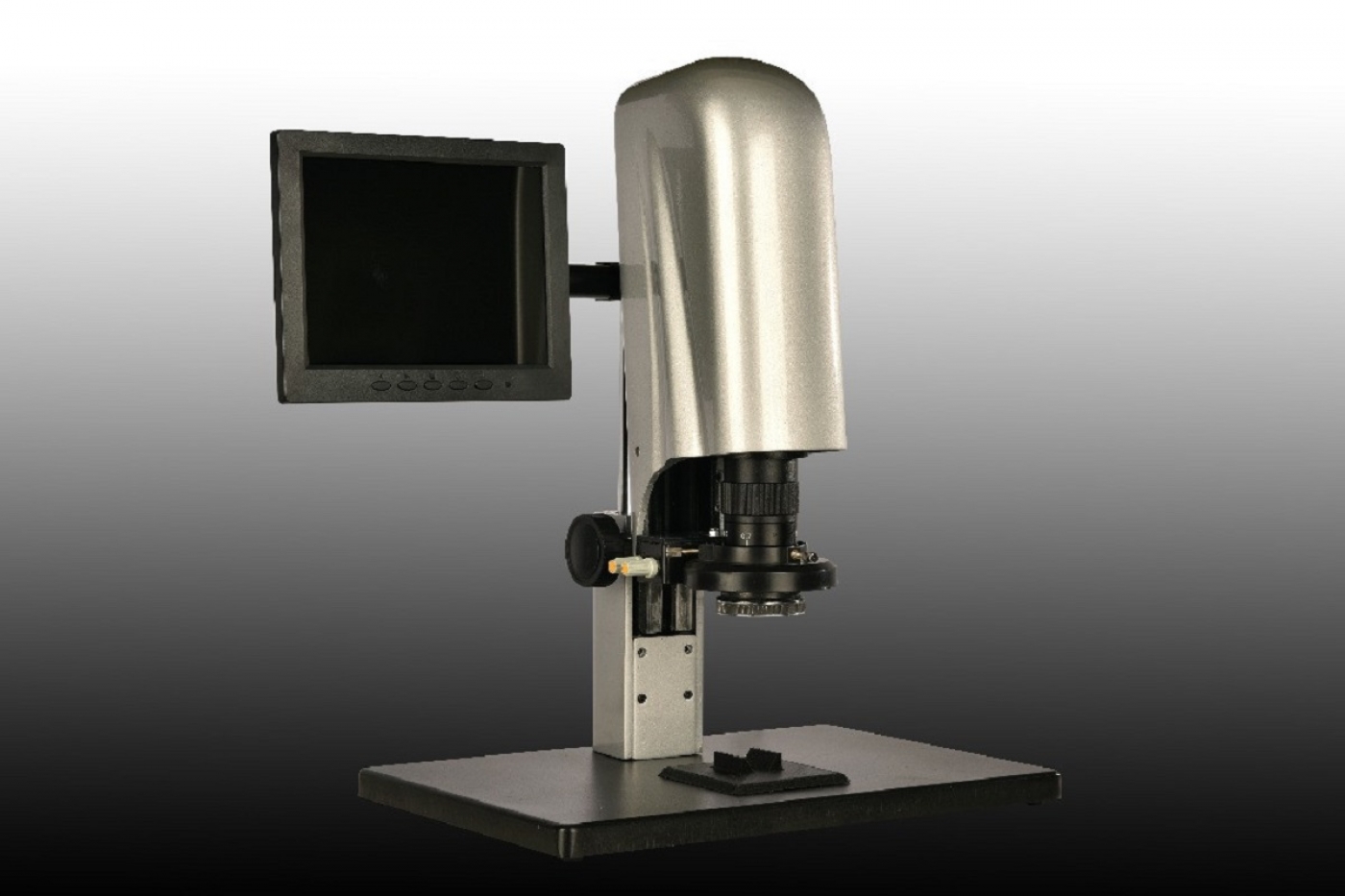
Fluorescence Molecular Imaging Systems were firstly introduced by Garofalakis and his colleagues. The first generation of this imaging system was designed for Non-contact Fluorescence Molecular Tomography. The molecular imaging system is a novel diagnostic method which is widely used in medicine. In the first generation, continuous argon laser was used to stimulate the fluorescent probes. In this system, a movable system scans the light points along the sample surface in the x and y directions. The sample stage can be moved using a rail along the camera optical axis. A CCD camera equipped with a cooling system receives the signal. The CCD camera is equipped with an objective lens which can be focused onto the specified area. In this special arrangement, to record different scenes, the laser source and the specimen are moveable, while the camera is fixed in its place. This imaging system can operate in both transillumination and epi-illumination modes. The way of selecting mode depends on the relative position of the device movable bases with light alignment tools for scanning. These movable bases can be placed on the camera’s side (in epi-illumination mode) or on the camera's optical axis (in transillumination mode). Finally, the data are processed by computer.
In the second generation, the laser beam goes through a beam scanner after passing through the telecentric scan lens. The scanner is equipped with galvanometer-controlled mirrors which are placed on highly sensitive engines. These engines change the mirrors direction in order for laser beam to move at two directions. The laser beam hits the mirror after passing through the scanner. After reflection of the beam from the first mirror, the beam is conducted by a second mirror towards the specimen. The second mirror is fixed above the system framework, while the first mirror is mounted on an optical table and moves between two fixed locations onto an optical rail.
The current imaging device, called Fluovision, is a non-invasive method, utilizing non-ionizing radiation and many clinical applications. This imaging technique is used in detection of cancer cells in molecular level. Currently, the most important application of this method is diagnosis of the development of the growth of breast and skin tumors. In addition, this imaging system is now used in imaging of lymph node map during operation. In this technique, the region is marked with a fluorescent substance and then subjected to the laser beam. The stimulated region emits non-uniform fluorescent beams according to Stokes shift. Fluorescent beams are captured with a highly sensitive CCD camera. In the fluorescent imaging method, the laser source and the detector are on the same side of the sample. This method is called epi-illumination mode. In this mode, fluorescent beam is collected from the tissue surface by a CCD camera.


 1871
1871 0
0  Printable